Micro Analysis and Visualization Lab (MAVL)
Lab Description
The Micro Analysis & Visualization Lab (MAVL) is an Institute-wide facility for microscopic analysis and imaging services. The MAVL provides researchers with access to a range of microscopic platforms including low-magnification stereozoom reflected-light (dissecting) scopes, transmitted-light compound scopes in a variety of configurations (including petrographic and fluorescence capabilities), inverted scopes for live cell or tissue analysis, and a high-magnification tabletop scanning electron microscope equipped for elemental composition analysis. Microscope cameras and basic image analysis software are available for many of the platforms. The MAVL was created in 2015 as a dedicated research space, equipment library and teaching/training facility that all at DRI can use, contribute to and share.
Equipment from the MAVL can be used by DRI researchers for projects such as characterization of pollutants in air and water samples, particle morphology and elemental analysis, identification of microfossils, or forensics. To discuss research needs related to micro-imaging and analysis, contact lab manager Dave Rhode at dave.rhode@dri.edu.
The MAVL provides researchers with access to a range of microscopic platforms, cameras and other equipment, including:
- Perkins-Elmer Opera imaging system – An automated high-content, high-throughput laser scanning confocal microplate screening and imaging system.
- Hitachi TM-1000 tabletop scanning microscope – An easy-to-use, high magnification scanning microscope that requires very little sample preparation. Has energy-dispersive x-ray spectroscopy capabilities.
- Olympus BX-51 transmitted light microscope – An all-around excellent general scope with Brightfield, phase contrast, and DIC objectives; basic polarizing capabilities; side port for training/teaching applications; and, digital camera and basic image measurement software package. Excellent for pollen, starch, phytoliths.
- Olympus BX-60 – a fluorescence microscope with a digital camera
- AmScope BH-200 – a fluorescence microscope with digital camera and basic image measurement package
- Arcturus Laser Capture and Microdissection System – for isolating and capturing specific cell samples from mixed cell populations for biomolecular analysis
- Nikon Eclipse TE-2000-U Inverted Live Cell Microscope
- Nikon Eclipse TE-300 Inverted Microscope with Eppendorf Micromanipulator System – has micromanipulator probes for efficient examination of tissues
- Microm HM 500 – Cryostat microtome for creating thin sections
- PCR-capable laminar flow cabinet for dust-free slide and sample preparation
- Rockhound – Hand-held portable Raman spectrometer
- Thermo-Niton XL3t GOLDD+ – Portable x-ray fluorescence spectrometer
- Nikon Optiphot and Labophot Optical Microscopes
- Stereozoom (Dissecting) Scopes
- Computers, monitors, basic image analysis software
- Digital and film cameras
- Objectives, condensers, stages, other spare parts
- Lamps
Training on some of the equipment is available upon request. Sample processing may be available on a case-by-case basis.
Dave Rhode, Ph.D.
Director of the Micro Analysis and Visualization Laboratory
Research Professor Archaeology
Division of Earth and Ecosystem Sciences
Contact: Dave.Rhode@dri.edu
CV/Bio: Dr. Dave Rhode
Dr. Rhode is a prehistorian, archaeobotanist, and paleoecologist with 40 years experience throughout western North America. His main focus of research concerns prehistoric human adaptations and paleoenvironmental change in arid environments. Towards this end, he has analyzed packrat middens and pollen from various paleoenvironmental locales, examined plant remains from various archaeological sites throughout western North America, and studied starch granules and pollenfrom archaeological sites and artifacts. In addition to his paleoenvironmental research, Dr. Rhode has directed several large archaeological programs for the US Department of Defense and Department of Energy. Since 2001, he has also developed a research program in the Tibetan Plateau of western China with colleagues Jeff Brantingham (UCLA), David Madsen (U Texas), John Olsen (U Arizona) and Charles Perreault (ASU).
Photo Gallery
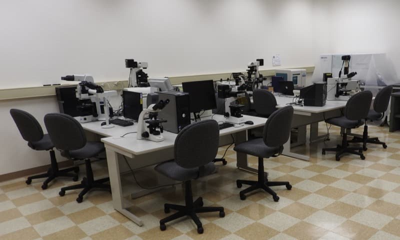
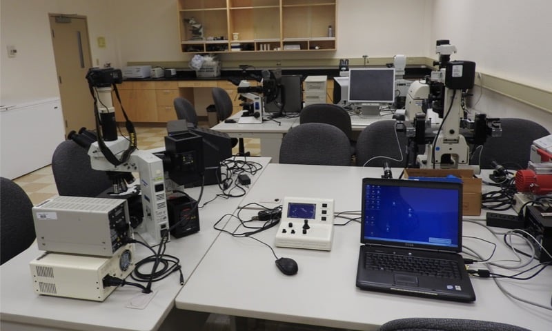
MAVL-workstation
Work stations in the MAVL can be configured to your specialized needs.
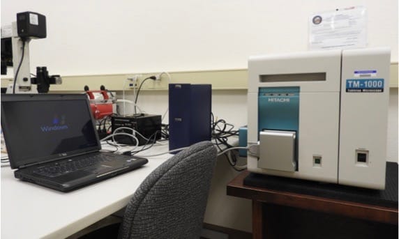
MAVL-hitachitm-1000
Hitachi TM-1000 tabletop scanning electron microscope, capable of 25-10,000X magnification. This unit is equipped with a Swift energy dispersive spectroscopic unit for elemental micro-characterization.

MAVL-willow-charcoal
An image obtained from the Hitachi TM-1000: Willow charcoal.
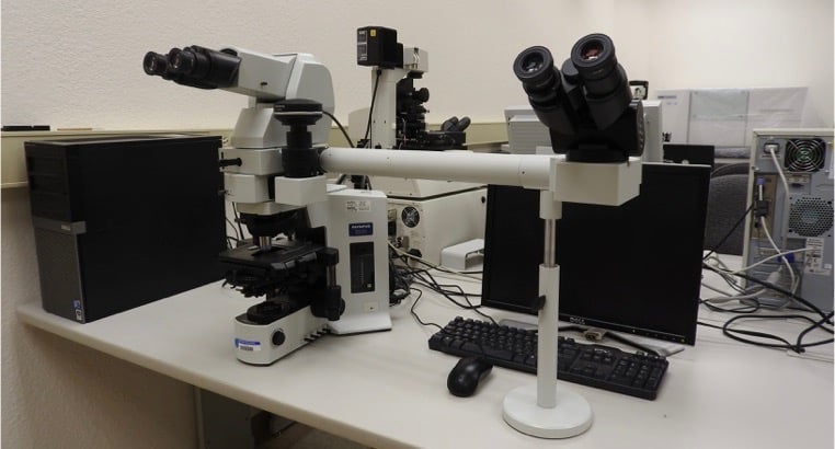
MAVL-olympus-bx51
Olympus BX-51 transmitted compound microscope, with side viewing attachment for teaching
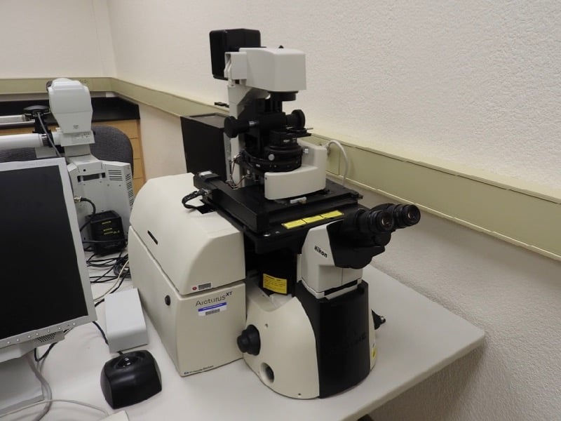
MAVL-nikon-eclipse-arcturis-laser
Nikon Eclipse inverted scope equipped with Arcturus laser microdissection system for cell capture and analysis.
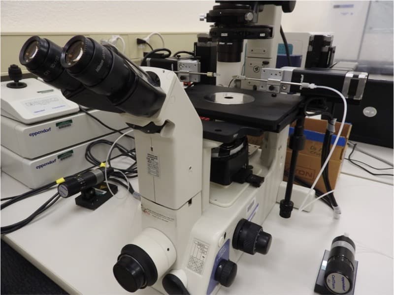
MAVL-nikon-eclipse-eppendorf-micromanipulators
Nikon Eclipse inverted scope equipped with Eppendorf micromanipulators for live cell/tissue sample examination.
MAVL-maxey-science-building
The MAVL is located in DRI’s Maxey Science Building, Room 148.
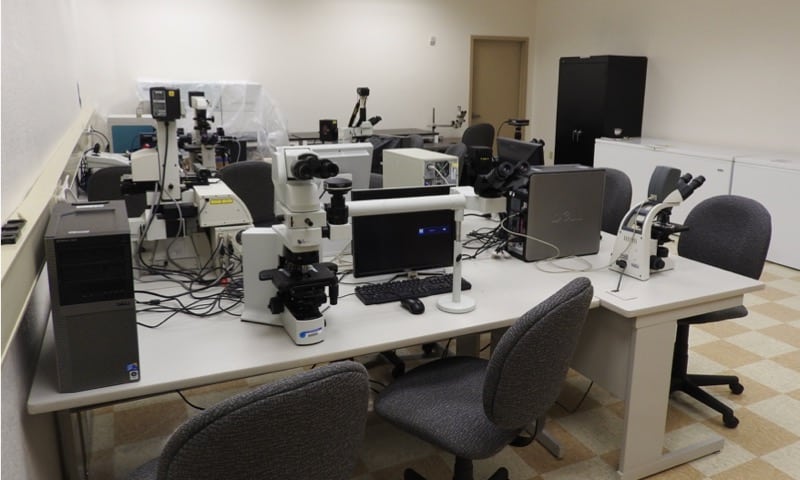
MAVL-equipment
To use the MAVL or to learn more about the equipment, please contact Dave Rhode.
CONTACT
Dave Rhode, Ph.D.
Lab Director
Dave.Rhode@dri.edu
LAB LOCATION
Desert Research Institute
2215 Raggio Parkway
Reno, NV 89512
DIVISION
Earth & Ecosystem Sciences